
NF1-Associated Plexiform Neurofibroma

Sunshine4CancerKids
Chaitanya Koduri, Ishanth Shantmoorthy, Eshal Fatima
July 20th, 2026
Abstract
Neurofibromatosis Type 1 (NF1) is a common inherited genetic disorder caused by pathogenic variants in the NF1 gene, which encodes the tumor suppressor protein neurofibromin and regulates the RAS/MAPK signaling pathway involved in cellular growth and proliferation (PubMed, 2013). Among the various manifestations of NF1, NF1-associated plexiform neurofibroma, a benign peripheral nerve sheath tumour, occurs in approximately 30-50% of individuals with NF1 and can cause chronic pain, neurological impairment, functional disability, and, in some cases, malignant transformation into malignant peripheral nerve sheath tumours (MPNSTs)(PubMed, 2025). This study explores how variation in access to diagnostic tools, including genetic testing, magnetic resonance imaging (MRI), and ultrasound, impacts neurodevelopmental outcomes and quality of life in affected children across the world. Additionlay, diagnosis of NF1-associated plexiform neurofibromas relies on clinical evaluation, genetic testing, advanced imaging techniques, and referrals from specialists for accurate tumor detection. However, unequal access to these resources in healthcare systems contributes to delayed diagnosis and poorer long-term outcomes. Specifically, delayed diagnosis of NF1-associated plexiform neurofibromas may allow tumors to progress during critical states of childhood growth and affect neurological development, increasing the risk of chronic pain, functional impairment, and diminished quality of life (World Health Organization, 2020). Overall, this study underscores the essential role of equal access to diagnostic tools, including genetic testing, MRI, and ultrasound, in early identification and clinical management of NF1- associated plexiform neurofibromas, as it highlights the importances of improving diagnostic tools accessibility and promoting early intervention, as its essential in ensurig that children with Koduri, Shantmoorthy, Fatima 3 NF1-associated plexiform neurofibromas receive appropriate care regardless of a countries economy and geographic location.
Discussion
Choosing to research NF1-associated plexiform neurofibromas was driven by our interest in understanding how access to healthcare can influence the lives of children with a genetic disorder. While advances in molecular biology and medical imaging have improved the diagnosis and management of NF1, our research revealed that these advances are not equally available around the world. This led us to investigate how differences in diagnostic tools affect the timing of diagnosing children affected by NF1-associated plexiform neurofibromas. Throughout this research, one of the most impactful ideas we discovered was that advances in medicine are only effective when patients have access to them. Although diagnostic technologies continue to improve, many children from third-world countries with limited resources still experience delays in early detection because these resources are not equally available. Ultimately, through this paper, we hope to encourage a greater understanding of the challenges faced by children living with NF1-associated plexiform neurofibromas and the importance of considering both scientific and social factors when addressing complex genetic disorders. Investigating this topic also demonstrated that improving patient outcomes depends not only on advances in medicine but also on recognizing the broader barriers that can limit access to care, as we overall hope that this paper encourages continued research into reducing healthcare inequality and inspires future efforts in making diagnosis and treatment more accessible to children all around the world.
Introduction
NF1-Neurofibroma is a cancer formed from the non-myelinating Schwann cells–fibroblasts–of the peripheral nervous system, primarily manifesting in early childhood and growing in severity with age. This benign cancer arises from a mutation in chromosome 17, which removes the mitotic safeguards of fibroblasts and facilitates rampant cell division. The excess growth of these cells eventually leads to the formation of large masses that infiltrate nerve tissue, causing tissue damage and dysfunction. Tumor growth typically occurs along nerve fibers, producing skin nodules and freckling. Children age six to ten are typically most effected, for while symptoms manifest between birth to age five, their severity peaks in late childhood (NF1 2026). However, current research has yielded solutions that may prove to be less invasive and more effective than current treatments. By targeting the specific pathways needed for cancer growth, MEK inhibitors produce lasting relief, cause less tissue damage, and require less hospitalization than conventional treatments, improving patient quality of life (MEK inhibitors 2026). However, despite advances in medical technology and tumor understanding, access is still limited based on wealth and technological advancement. As a result, geographic barriers to treatment are formed, preventing patients of poorer geographic regions from receiving the care they need. In this landscape of hurdles, the neurodevelopmental outcomes of patients with NF1-Associated Plexiform Neurofibroma vary drastically based on geographic location. Thus, with this disparity, the accessibility of a treatment is more crucial than its effectiveness. By improving awareness of NF1 and its underlying genetic causes, patient outcomes may be improved drastically and the geographic gap overcome.
Disease Background and Biology
NF1-associated plexiform neurofibromas (PNs) are a type of benign peripheral nerve sheath tumors that develop in individuals with Neurofibromatosis Type 1 (NF1), an inherited genetic disorder caused by pathogenic variants in the NF1 gene located on chromosome 17 (Journal of Medical Genetics, 2008). NF1 follows an autosomal dominant pattern of inheritance, which means that only one altered copy of the NF1 gene is needed for the disorder to occur. Although approximately 50% of cases are inherited from an affected parent, the remaining cases result from spontaneous mutations that take place during early fetal development(Cell Press, 2014). Additionally, plexiform neurofibromas are benign, as they can grow extensively along the peripheral nerves, causing pain, neurological impairment, and disability. Specifically, in about 8-15% of cases, these tumors may transform into malignant peripheral nerve sheath tumors, overall highlighting the importance of long-term monitoring and early diagnosis in accurately managing the tumor's growth
.

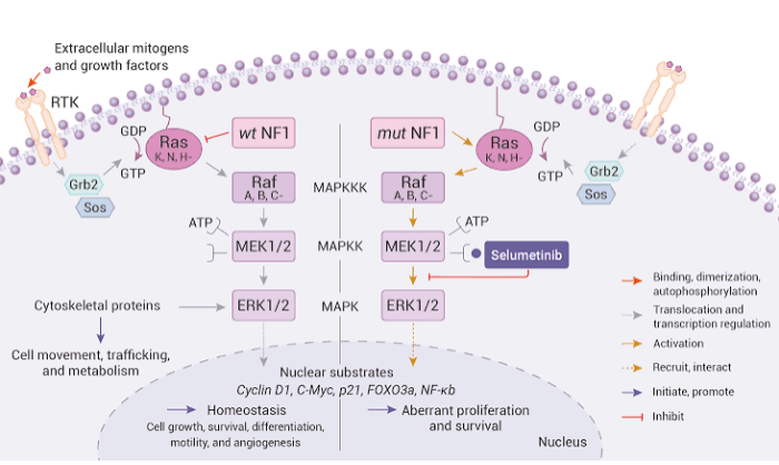
Furthermore, the development of NF1 is directly linked to the loss of neurofibromin, a tumor suppressor protein encoded by the NF1 gene, helping regulate the RAS/MAPK signaling pathway, which controls cell growth and differentiation (American Association for Cancer Research, 2011). Under normal conditions, the neurofibromin inactivates the RAS protein after it has transmitted a growth signal, preventing unnecessary cell division. However, when mutations reduce the production of neurofibromin, RAS remains continuously active, causing constant activation of downstream signaling proteins within the pathway. As a result, the cells continue to proliferate even after growth signals have stopped, consequently creating the conditions necessary for tumor development
Furthermore, although every human with NF1 carries one mutated copy of the NF1 gene, plexiform neurofibromas generally form only after a second mutation occurs within the Schwann cells, during a process called the two-hit hypothesis (American Association for Cancer Research, 2011). Schwann cells normally produce the myelin sheath surrounding peripheral nerves, allowing for electrical impulses to travel efficiently throughout the nervous system, and in the case of NF1, a complete loss of neurofibromin causes the cells to proliferate uncontrollably while simultaneously recruiting blood vessels, mast cells, macrophages, and fibroblasts to help fight the tumor. Consequently, these cells create a complex tumor microenvironment that supports continued tumor growth and contributes to the creation of the structural characteristics of plexiform neurofibromas (Neurofibromatosis Therapeutic Acceleration Program, 2024).

Unlike many other benign tumors, plexiform neurofibromas grow diffusely along multiple branches of peripheral nerves rather than remaining confined to a single location. This infiltrative growth pattern makes complete surgical removal difficult without damaging the healthy nerve tissues ( Society for NeuroOncology, 2022). Therefore, understanding the molecular biology of NF1 provides important insight into how mutations in the NF1 gene disrupt normal cellular regulation and lead to the development of plexiform neurofibromas in children. Overall, this biological foundation is essential for understanding the methods used to diagnose these tumors, as the knowledge of their genetic and cellular components helps guide proper diagnosis.
Diagnosis
Diagnosing Neurofibromatosis Type 1 (NF1), the underlying disorder associated with plexiform neurofibromas, is commonly based on the diagnostic criteria of the National Institutes of Health(NIH). Specifically, physicians evaluate patients for characteristic features, including cafe-au-lait macules, axillary and inguinal freckling, optic pathway gliomas, plexiform neurofibroma, distinctive osseous lesions, and a confirmed pathogenic variant of the NF1 gene (Neurosurgery, 2021). Additionally, since many of these features develop gradually throughout childhood, younger children may not initially meet all the criteria, resulting in continued clinical follow-up. Although NF1 may be suspected during infancy because of the presence of café-au-lait macules, plexiform neurofibromas is most commonly identified during early childhood and later diagnosed before ten years old (Children’s Tumor Foundation, 2026). The first signs of a plexiform neurofibroma often include a soft tissue mass beneath the skin, localized swelling, chronic pain, numbness, muscle weakness, and reduced range of motion (American Journal of Medical Genetics, 2022).
Following the initial physical examination, imaging also plays a critical role in confirming the diagnosis and determining the extent of tumor growth. For example, Magnetic resonance imaging (MRI) is considered the gold standard in the evaluation of plexiform neurofibromas because it provides highly detailed images of soft tissues, peripheral nerves, and surrounding anatomical structures without exposing patients to ionizing radiation (Journal of Medical Genetics, 2008). Moreover, it also allows physicians to assess tumor size and location accurately. Another diagnostic tool used to assess NF1 would be an ultrasound, which is used to evaluate superficial plexiform neurofibromas, as it is non-invasive, widely available, and does not require radiation exposure (Clinical Neurophysiology,2018). However, an ultrasound cannot Koduri, Shantmoorthy, Fatima 10 provide the same level of detail as the MRI and is generally used as a complementary imaging technique in diagnosing NF1-associated plexiform neurofibromas.

In addition to imaging, another diagnostic tool used is genetic testing, which confirms pathogenic variants in the NF1 gene, especially in children who have not yet developed enough clinical features to satisfy the NIH diagnostic criteria (Journal of Neuroadiology, 2019). Once a diagnosis has been established, patients are referred to different types of specialists, depending on the location of the tumor and the severity of clinical symptoms. Furthermore, unlike many cancers, plexiform neurofibromas are not staged using the traditional TNM classification system of Tumor, Node, Metastasis because they are benign (Wiley Online Library, 2009). Hence, physcians evalluate its reiks based on the symptoms, tumor size, and anatomical location, and potential complications on other vital organs. Overall, diagnosing NF1-associated plexiform neurofibromas requires combining evaluations, examinations, genetic testing, imaging techniques, and specialist referrals to accurately identify the disease and monitor the tumor's progression. Once a diagnosis has been confirmed, healthcare providers can then determine the most effective treatment approach based on the patient's individual needs and characteristics of the tumor. Therefore, understanding the diagnostic process provided the foundation for examining the current standard of care used to manage NF1-associated plexiform neurofibromas.
Current Standard of Care
After diagnosis, plexiform neurofibroma can be treated in several ways depending on the size and location of the tumor. The chief means of treatment is surgical removal, wherein large or compromising growth is excised. To prevent regrowth, this is often supplemented with adjuvant anthracycline-based chemotherapy and localized doses of radiation. This cocktail of treatments targets cell DNA replication mechanisms, thereby removing the ability of tumor cells to multiply. Through this means of treatment, tumor mass is reduced significantly, thereby preventing growth into malignancy. Yet, surgery is limited due to the proximity of many neurofibromas to sensitive nerve tissue or organs. (Chemotherapy Treatment 2013). As roughly 35% of pediatric patients fulfill the aforementioned criteria, this method automatically excludes a significant group of patients (Clinical Characteristics and Management 2022) . Additionally, surgery does not prevent malignancy, for while it reduces tumor mass, it does not target the underlying genetic cause for growth.
As a result, the overall risk of malignancy for neurofibromas remains the same even after surgery. This means that while a potentially malignant tumor is removed, there is still a significant risk of a different tumor developing malignancy (NF1 2026). Surgery’s inherent invasiveness, furthermore, limits its continued use. Despite surgical intervention, tumor growth still continues, necessitating successive excisions. Potentially, continued surgery leads to tissue damage (Liao et al 2025). Eventually, some patients to grow malignant over time. In this case, survival rates are reduced significantly, with MPNST patients experiencing survival rates of 50-35% (Chemotherapy Treatment 2013).

This is compounded by the greater degree of tissue infiltration of MPNSTs compared to early stage neurofibromas, with roughly 8.3% of MPNST tumors found in pelvic bone and the thoracic cavity, areas that are harder to reach with surgery (Malignant Nerve Sheath Tumor 2023).

However, surgery, as stated earlier, is not a sole means of treatment. Oftentimes it is supplemented with chemotherapy to reduce tumor regrowth. However, chemotherapy is insufficient in this purpose. While chemotherapy can reduce tumor growth, it rarely stops it completely. In a study conducted by the National Institutes of Health, patients experienced continued tumor growth rates of 25-30% (Chemo Institutional Review 2013).
Chemotherapy additionally poses a threat to both healthy and cancerous tissue. By destroying the cellular machinery needed to multiply, anthracycline-based chemotherapy can disrupt the healthy function of tissue, leading to organ failure. Though impacting all rapidly multiplying cells, this is of particular concern in cardiac tissue, whose greater metabolic demands lead to the imbibing of greater quantities of anthracycline compounds. In turn, apoptosis (cell death) is induced, leading to the loss of cardiac function and death in a condition known as anthracycline based cardiac toxicity (NIH Library of Medicine N.D).
The effects of this are most keenly felt in children, as their tissues are more primed to react to apoptotic stress than adult tissues, resulting in greater susceptibility to chemotherapy-induced tissue damage (Mdege, 2016). For adjuvanant chemotherapies primarily work through induced cell death, their use may actually increase the risk of mortality in youth patients by damaging vital tissues an organ systems. Thus, the destructiveness of conventional chemotherapy, coupled with the limited application of surgical intervention, makes it unsuitable for use in pediatric patients with NF1. However, with the development of more targeted treatments, these hurdles will be overcome. Ultimately, the current treatments for neurofibroma provide bar by which future methods may be measured.
Treatments
There are two principal treatment routes that currently exist for managing NF1-Associated Plexiform Neurofibromas, and it’s useful to think of them as a blunt weapon versus a precision instrument, because the contrast explains almost everything about why access to each one matters so much.
Surgical resection, sometimes used alongside conventional chemotherapy in more aggressive cases, has long been the default standard of care. But surgery here behaves more nuke-like, detonating a blast radius around the target and less with the precision of a scalpel; because plexiform neurofibromas grow diffusely along and through nerve fibres rather than forming one clean, removable mass, surgeons often cannot take the tumour out without also damaging the nerve it has grown onto (NTAP, 2024). Complete resection is rarely achievable and even when it appears successful, recurrence is common. Every additional surgery raises the risk of permanent nerve damage and lost function, and (Liao et al.’s 2025) review notes that management of NF1 more broadly requires a whole-patient balancing act, given how the condition’s complications extend well beyond any single tumour to include skeletal abnormalities, epilepsy and other neurolodevelopmental involvement across the patient’s life. Conventional chemotherapy has similarly struggled to offer a durable solution; trials of cytotoxic agents such as vinblastine and methotrexate found that tumours largely remained stable at best, with none of the participants achieving an actual reduction in tumour volume, while nearly half showed measurable disease progression by the end of the treatment (Vinblastine/Methotrexate Phase 2 Study, MDPI, 2023). Beyond limited effectiveness, conventional chemotherapy has also been associated with substantial secondary toxicity, including damage to bone marrow function and the gastrointestinal system. Surgery itself is often impractical as a repeated solution, giventhe blood loss, extended recovery and physical toll involved in operating on tumours that are deeply intertwined with nerve tissue, particularly when multiple procedures are required over a patient’s lifetime. Rather than resolving the problem permanently by addressing the underlying biology, surgery frequently does little more than reset the tumour’s growth temporarily, with regrowth commonly being observed within a few months following each procedure. In essence, surgery removes tissue indiscriminately, taking healthy nerves along with diseased tissue simply because the two cannot be cleanly separated.
Selumetinib, by contrast, works much more with the precision of a scalpel operating at a molecular level. As an MEK inhibitor, it selectively blocks the MEK1 and MEK2 proteins that become hyperactive when the NF1 gene, neurofibromin, is faulty, interrupting the exact RAS/MAKP signalling pathway driving the tumour’s growth, rather than attempting to physically remove it. (Rare Disease Advisor, 2024). It is administered orally as a capsule, taken twice daily roughly twelve hours apart, with dosage individualised by body surface area rather than a flat dose. Koselugo, the brand name under which it is marketed, is only available through a single specialty pharmacy rather than being available at ordinary retail pharmacies (Alexion Access Navigator, 2026). In the SPRINT trial that supported its approval, around 70% of paediatric participants saw their target tumour shrink by at least 20% (PubMed, Neurofibromatosis Program, 2020), leading the FDA to approve it in 2020 for children aged two or older with inoperable NF1 Plexiform Neurofibromas - the first drug ever approved specifically for NF1 (PubMed, Neurofibromatosis Program, 2020). Approval has since broadened, initially to younger children in 2025 and then to adults in November 2025 following the KOMET trial (ASCO Publications, 2025). Selumetinib’s emergence marks a genuine turning point in NF1-PN management as it’s the first time clinicians have had a tool that addresses thetumour’s underlying biology rather than simply attempting to physically outmanoeuvre it. Ongoing trials into related MEK inhibitors, such as mirdametinib, suggest this is not an isolated breakthrough but the beginning of a broader shift toward targeted, mechanism-based therapy for a condition that, until recently, offered patients very few options besides surgery (MDPI Comprehensive Review, 2025).
Limitations
MEK inhibitors, despite their effectiveness, face significant barriers to accessibility. Chief among these are cost and distribution. Selumetnib, trademarked as “Koselugo” is the sole approved drug in its category. As such, Selumetnib distribution is limited to its proprietary manufacturer: Astrazeneca, preventing it from being sold in a cheaper generic form. This situation ensures that the demand constantly outpaces supply, maintaining astronomically high prices. As of 2026, the annual price for Koselugo treatment is $268,677, making it far out of reach of the average NF1 patient (Cost Comparison N.D). Furthermore, the corporate stranglehold over Koselugo production also restricts treatment distribution, with patients only able to access treatment through direct interaction with proprietary Astrazeneca software. As wealthier patients in first-world countries have the support systems and income necessary to access treatment, this system isolates poorer patients and those in 3rd world countries. Among first-world countries, the systems in place to receive treatment vary across the globe, with certain nations receiving more favorable policies than others. Due to the complex interplay between government and insurance, some geographic regions or countries may have inconsistent eligibility restrictions or no chance for reimbursement at all. This primarily impacts poorer and middle-class patients in first-world countries, for they are less able to afford the same degree of insurance protection as their wealthier counterparts. As such, they are more dependent on public support systems or cheaper insurance plans to afford treatment. In Slovakia, for example, only a minority of 19% of applicants receive publicly funded reimbursement for Koselugo treatment (T Hofmarcher, P Szilagyiova et. al N.D). Closer to home, Canadian ethics panels have proposed public “compassionate” access programs to facilitate drug distribution to poorer and underinsured patients (CADH Review N.D). On the other hand, the general lack of financial support systems in 3rd world countries excludes patients from poorer geographic regions such as Asia or Africa from care. In these areas, insurance is generally composed of private programs, keeping care out of the reach of most citizens. India, the most populous country in Asia, largely does not use public insurance, with its private programs being significantly more effective (Pros and cons of Health insurance Bajaj). As opposed to the general annual cap of public insurance of $5,000, private insurance can reimburse significantly larger sums of up to $104,000 (How Much Health Insurance do You Need? N.D). Nigeria, the most populous country in Africa, has a similar system, with public care covering small fees for laborers and private care for wealthier patients (National Health Insurance Authority N.D). Largely, the insurance systems of these countries are closely mirrored by the vast majority of nations in their respective regions. However, neither these countries nor those sharing their template have publicly funded access to Koselugo, barring a vast majority of their NF1 patient population from treatment. This is in part facilitated by the general economic inequality as a sizeable fraction of the populations of these regions work under informal systems, with roughly 68% (Informal Economy 2018) and Asian workforce and 85% of the African workforce employed informally (Informal Economy In Africa, 2022). Consequently, this large proportion is dependent on public support, preventing a majority of citizens from receiving NF1 treatment. Thus, the cost of Koselugo directly weighs into its accessibility, reducing the already weak viability of financial support structures in 3rd world countries and firmly placing vital treatment out of the hands of the vast majority of the global population. Ultimately, despite advances in research and technology, the chief limitation of treatment is not the disease itself, but the inequality in its access. Thus, the systems by which NF1 is treated form the base upon which treatment efficiency can be improved, ensuring equality among patients regardless of geographic locales.
Statistics
While neurofibromatosis type 1 is classified as a rare genetic disorder, its clinical relevance extends far beyond its prevalence numbers alone, owing to the wide variability in how the condition progresses across patients. NF1 arises from mutations in a single tumour-suppressor gene, yet affects roughly 1 in every 3,000 people worldwide, regardless of geography, ethnicity or sex, a rate that’s consistent enough that most general practitioners will encounter at least one NF1 patient over the course of their career (European Medical Journal, 2021). Among the many manifestations of NF1, plexiform neurofibromas represent one of its most clinically significant complications; between 30% to 50% of NF1 patients develop them, making PN less a rare complication and more like a coin-flip likelihood for any NF1 patient (European Medical Journal, 2021).
Diagnosis tends to come early rather than late, though estimates vary by setting, with a median age of PN diagnosis of around 5-8 years old, though onset is frequently recognised as early as the first year of life (Science Direct, 2025). A South China survey of 389 paediatric patients similarly found a median age of 6.1 years (Science Direct, 2026), while a South African cohort of 48 children with NF1 recorded a median presentation age of just 4 years (PubMed, South African NF1 phenotype study, 2006). Plexiform neurofibromas themselves behave unpredictably once formed. A retrospective cohort study tracking 201 NF1 patients through repeated whole-body MRI recorded a median tumour growth rate of 3.7% per year, though with substantial variation between individuals, and growth was consistently faster in younger patients than in older ones (PubMed, 2012). Roughly 13.5% of tumours grew significantly enough over the study period to be classified as a clinical concern, disproportionately amongst children with the tumour’s growth rate being correlated with age, thus reinforcing early childhood as the period during which surveillance matters most, and also the period in which it is least consistently delivered (PubMed, 2012). Malignant transformation represents the most serious, if less common, complication of plexiform neurofibromas, and it disproportionately drives NF1-related mortality despite only affecting a minority of the patients. Multiple independent cohort studies converge on a lifetime risk of roughly 8-13% for progression to malignant peripheral nerve sheath tumour, or MPNST, which almost always arises from a pre-existing plexiform neurofibroma rather than developing independently (Oxford Academic Neuro-oncology Advances, 2025). In one large cohort of 1,067 adult NF1 patients, plexiform neurofibromas were present in 47% of cases and while overall mortality within the group was relatively low at 5%, MPNST was identified as the most common cause of death amongst those who died, with a median age of just 48 years (Oxford Academic Neuro-oncology Advances, 2025). The same study noted that surgical management of PNs was frequently limited by regrowth and complications, reinforcing why a targeted drug treatment such as selumetinib, rather than surgery alone, matters most for this kind of highest-risk group in which access to that same treatment is often least available.
Impacts
A plexiform neurofibroma diagnosis rarely stays contained to the tumour itself. Children with NF1-PN commonly live with pain that interrupts school and play, visible disfigurement that shapes how peers treat them and for a relative proportion as well as motor difficulty. One USA survey of patients and caregivers found pain in 64.6% of cases, disfigurement in 32.9% and motor dysfunction in nearly a third, often accompanied by repeated debulking surgeries through childhood (PubMed, 2022). These are not background symptoms; they surface during the exact years a child is first learning who they are and how the world responds to them. This burden is not experienced equally across healthcare systems and it connects directly to the disparities in diagnostic access discussed earlier in this paper. In well-resourced areas, a 6.1 year median diagnosis age likely already reflects a level of monitoring through regular paediatric reviews, accessible imaging, and specialist referral pathways that already exist and are used.
This is far from guaranteed in lower-income settings, where the same systemic barriers already identified (limited MRI availability, scarce genetic testing infrastructure and a shortage of specialists trained to recognise early NF1 presentation) mean this gap between onset and diagnosis is likely to stretch considerably longer. Evidence suggests that even a comparable or earlier presentation age offers no guarantee of comparable outcomes; school-related learning and behavioural problems were documented in 70% of school-aged children with NF1 in a South African cohort, compared to 29.8-45% reported in international literature, despite the median presentation age of just 4 years old (PubMed, South African NF1 phenotype study, 2006), an early sign that when a child is seen matters less than what happens for them afterwards. Since psychological and developmental support pathways are frequently only triggered once a formal NF1 diagnosis is made, a delayed or under-supported diagnosis does not only postpone treatmentfor the tumour itself, it also postpones access to the psychological and neurodevelopmental support increasingly recognised as necessary for NF1 paediatric patients, given the condition’s well-documented association with learning difficulties, attention difficulties and reduced quality of life. Visible tumours are linked to reduced self-esteem and increased likelihood of being singled out or bullied, and NF1 more broadly carries elevated rates of anxiety and depression compared to the general population (PubMed, 2023). Despite the psychological toll frequently outlasting the physical one, psychological support is rarely built into standard care as a default; where it exists, it depends heavily on whether a given clinic happens to have the resources to offer it.
In this sense, the gap between onset and diagnosis is of paramount importance: it is one of the clearest points at which global inequities in healthcare access translate directly into unequal developmental and psychological outcomes for children with the same underlying genetic condition worldwide.
Conclusion
In the current landscape for NF1 patients, both treatment and awareness remain unequal based on geographic location and socioeconomic factors. Despite the advent of more effective treatment methods, this problem has only been exacerbated. However, the rampant inequality facing NF1 patients in poorer nations can be remedied by promoting awareness and disseminating more readily accessible tools for diagnosis. Through advocacy programs, understanding of tumor symptoms can be improved in isolated geographic locales and diagnosis rates can be improved. This can be supplemented with the use of technology and psychology to further pinpoint at-risk individuals in populations. By using this approach in foreign aid, patients may be given effective preliminary support and quality of life may be improved. Yet, many challenges still remain. Chiefly, the ineffective support systems of the 3rd world and the high cost of new proprietary treatments keep vital care out of the hands of a large fraction of patients. In this bright new dawn for NF1 treatment, accessibility and understanding are key to improving patient quality of life. Research must be conducted to gain a superior understanding of the means by which tumors multiply and spread to create newer treatments free of proprietary restrictions to allow all patients to afford cutting-edge treatment. Ultimately, care quality can be standardized between geographic regions through this combination of human ingenuity and spirit, allowing bright “rays” of hope to fall on patients all over the world.
Bibliography (MLA)
1. Children's Tumor Foundation. Children's Tumor Foundation. Children's Tumor Foundation, https://www.ctf.org/. Accessed 22 July 2026. 2. National Center for Biotechnology Information. GeneReviews®: Neurofibromatosis 1. National Library of Medicine, https://www.ncbi.nlm.nih.gov/books/NBK1109/. Accessed 22 July 2026. 3. World Health Organization. World Health Organization. World Health Organization, https://www.who.int/. Accessed 22 July 2026. 4. Neurofibromatosis Therapeutic Acceleration Program. Neurofibromatosis Therapeutic Acceleration Program, 2024, n-tap.org. Accessed 23 July 2026. 5. Group of Neurofibromatosis Plastic and Reconstructive Surgery Society of Chinese Medical Association, et al. “Interpretation of the Expert Consensus for Full Course Management of Plexiform Neurofibroma (2025 Edition).” Zhongguo Xiu Fu Chong Jian Wai Ke Za Zhi, 2026, PubMed, https://pubmed.ncbi.nlm.nih.gov/41981422/. Accessed 23 July 2026. 6. Riccardi, V. M. “A Controlled Multiphase Trial of Ketotifen to Minimize Neurofibroma-Associated Pain and Itching.” Archives of Dermatology, vol. 129, no. 5, 1993, pp. 577–581. PubMed, https://pubmed.ncbi.nlm.nih.gov/8481017/. Accessed 23 July 2026. 7. “Advances in Therapy for Patients with NF1: Selumetinib and MEK Inhibition.” Rare Disease Advisor, 2024, https://www.rarediseaseadvisor.com/insights/advances-therapy-patients-nf1-selumetinibmek-inhibition/. Accessed 23 July 2026. 8. Gross, Andrea M., et al. “Selumetinib for Children with Neurofibromatosis Type 1 and Plexiform Neurofibromas: A Plain Language Summary of SPRINT.” Future Oncology, vol. 20, no. 14, 2024, pp. 877–890. PubMed, https://pubmed.ncbi.nlm.nih.gov/38869947/. Accessed 23 July 2026. 9. Chen, Alice P., et al. “Final Analysis of KOMET (NCT04924608), a Phase 3 Study of Selumetinib in Adults with NF1-PN.” Journal of Clinical Oncology, vol. 44, no. 16_suppl, 2026, p. 3110. American Society of Clinical Oncology (ASCO) Annual Meeting Abstract, https://doi.org/10.1200/JCO.2026.44.16_suppl.3110. Accessed 23 July 2026. 10. Author(s). “Article Title.” Journal of Clinical Medicine, vol. 14, no. 14, 2025, article 5071. MDPI, https://www.mdpi.com/2077-0383/14/14/5071. Accessed 23 July 2026. 11. Author(s). “Article Title.” Cancers, vol. 15, no. 9, 2023, article 2621. MDPI, https://www.mdpi.com/2072-6694/15/9/2621. Accessed 23 July 2026. Koduri, Shantmoorthy, Fatima 27 12. How to Access Koselugo. Alexion Access Navigator, 2023, https://alexionaccessnavigator.com/-/media/alexionaccessnavigator/koselugo/files/how-to -access-koselugo-brochure-(2).pdf. Accessed 25 July 2026. 13. Tucker, T, et al. “Longitudinal Study of Neurofibromatosis 1 Associated Plexiform Neurofibromas.” Journal of Medical Genetics, vol. 46, no. 2, BMJ, Oct. 2008, pp. 81–85, https://doi.org/10.1136/jmg.2008.061051. Accessed 24 July 2026. 14. Chen, Zhiguo, et al. “Cells of Origin in the Embryonic Nerve Roots for NF1-Associated Plexiform Neurofibroma.” Cancer Cell, vol. 26, no. 5, Cell Press, Oct. 2014, pp. 695–706, https://doi.org/10.1016/j.ccell.2014.09.009. Accessed 24 July 2026. 15. Le, Lu Q., et al. “Susceptible Stages in Schwann Cells for NF1-Associated Plexiform Neurofibroma Development.” Cancer Research, vol. 71, no. 13, American Association for Cancer Research (AACR), June 2011, pp. 4686–95, https://doi.org/10.1158/0008-5472.can-10-4577. Accessed 24 July 2026. 16. Fisher, Michael J, et al. “Management of Neurofibromatosis Type 1-associated Plexiform Neurofibromas.” Neuro-Oncology, vol. 24, no. 11, June 2022, pp. 1827–44, https://doi.org/10.1093/neuonc/noac146. Accessed 24 July 2026. 17. Belakhoua, Sarra M., and Fausto J. Rodriguez. "Diagnostic pathology of tumors of peripheral nerve." Neurosurgery 88.3 (2021): 443-456. Accessed 25 July 2026. 18. “Neurofibromatosis Type 1: Burden of Disease in Patients with Plexiform Neurofibromas.” European Medical Journal (EMJ Reviews), Apr. 2021, https://www.emjreviews.com/oncology/symposium/neurofibromatosis-type-1-burden-ofdisease-in-patients-with-plexiform-neurofibromas-j190421/. Accessed 25 July 2026. 19. Longo, Jody F., et al. “Recent Advances in the Diagnosis and Pathogenesis of Neurofibromatosis Type 1 (NF1)-Associated Peripheral Nervous System Neoplasms.” Advances in Anatomic Pathology, vol. 25, no. 5, Ovid Technologies (Wolters Kluwer Health), Sept. 2018, pp. 353–68, https://doi.org/10.1097/pap.0000000000000197. Accessed 25 July 2026. 20. Uthoff, J., et al. “Radiomic Biomarkers Informative of Cancerous Transformation in Neurofibromatosis-1 Plexiform Tumors.” Journal of Neuroradiology, vol. 46, no. 3, Elsevier Masson, June 2018, pp. 179–85, https://doi.org/10.1016/j.neurad.2018.05.006. Accessed 25 July 2026. 21. Telleman, Johan A., et al. “Nerve Ultrasound in Neurofibromatosis Type 1: A Follow-Up Study.” Clinical Neurophysiology, vol. 129, no. 2, Elsevier BV, Feb. 2018, pp. 354–59, https://doi.org/10.1016/j.clinph.2017.11.014. Accessed 25 July 2026. 22. Sanefuji, Masafumi, et al. “Clinical Characteristics and Management of Plexiform Neurofibromas in Children with Neurofibromatosis 1: A Japanese Nationwide Survey.” Brain and Development, vol. 47, no. 1, 2025, article 104300. Elsevier, https://doi.org/10.1016/j.braindev.2024.10.008. Accessed 25 July 2026. 23. Porter, D. E., et al. “Survival in Malignant Peripheral Nerve Sheath Tumours: A Comparison between Sporadic and Neurofibromatosis Type 1-Associated Tumours.” Koduri, Shantmoorthy, Fatima 28 Sarcoma, vol. 2009, 2009, pp. 1–5, https://doi.org/10.1155/2009/756395. Accessed 25 July 2026. 24. Morris, Stephanie M., et al. “Predictive Modeling for Clinical Features Associated with Neurofibromatosis Type 1.” Neurology: Clinical Practice, Apr. 2021, p. 10.1212/CPJ.0000000000001089, https://doi.org/10.1212/cpj.0000000000001089. Accessed 25 July 2026. 25. Nguyen, Rosa, et al. “Growth Dynamics of Plexiform Neurofibromas: A Retrospective Cohort Study of 201 Patients with Neurofibromatosis 1.” Orphanet Journal of Rare Diseases, vol. 7, 2012, article 75. https://pubmed.ncbi.nlm.nih.gov/23035791/. Accessed 26 July 2026. 26. Salamon, Johannes, et al. “Malignant Peripheral Nerve Sheath Tumors in Neurofibromatosis Type 1 Arise from Distinct Nodular Lesions: A Retrospective Imaging Analysis.” Neuro-Oncology Advances, vol. 7, no. 1, 2025, article vdaf201. Oxford University Press, https://academic.oup.com/noa/article/7/1/vdaf201/8253364. Accessed 26 July 2026. 27. Yi, Meng, et al. “Clinical Characteristics of 389 Pediatric Patients With Neurofibromatosis Type 1 in South China.” Pediatric Neurology, vol. 181, 2026, pp. 107–113. Elsevier, https://www.sciencedirect.com/science/article/pii/S0887899426001669. Accessed 26 July 2026. 28. Tucker, T., et al. “The Natural History of Internal Plexiform Neurofibromas in Neurofibromatosis 1.” Orphanet Journal of Rare Diseases, vol. 1, 2006, article 13. https://pubmed.ncbi.nlm.nih.gov/16551456/. Accessed 26 July 2026. 29. Yang, Xiaoqin, et al. “Clinical and Humanistic Burden among Pediatric Patients with Neurofibromatosis Type 1 and Plexiform Neurofibroma in the USA.” Child's Nervous System, vol. 38, no. 8, 2022, pp. 1513–1522. https://pmc.ncbi.nlm.nih.gov/articles/PMC9325812/. Accessed 26 July 2026. 30. Cavallo, Nicola Davide, et al. “Neuropsychiatric Manifestations, Reduced Self-Esteem and Poor Quality of Life in Children and Adolescents with Neurofibromatosis Type 1 (NF1): The Impact of Symptom Visibility and Bullying Behavior.” Children, vol. 10, no. 2, 2023, article 330. https://pmc.ncbi.nlm.nih.gov/articles/PMC9955783/. Accessed 26 July 2026. 31. "Alexion Navigation." Alexion, Astrazeneca, alexionaccessnavigator.com/-/media/alexionaccessnavigator/koselugo/files/how-to-access -koselugo-brochure-(2).pdf. 32. "Anthracycline Toxicity." National Library of Medicine, National Institute of Health, www.ncbi.nlm.nih.gov/books/NBK599501/. 33. "CADTH Reimbursement Review." Canjhealthtechnol.ca, canjhealthtechnol.ca/index.php/cjht/article/view/SR0764r/1477. Koduri, Shantmoorthy, Fatima 29 34. "Chemotherapy for the treatment of malignant peripheral nerve sheath tumors in neurofibromatosis 1: a 10-year institutional review." Nih.org, National Institute of Health, pmc.ncbi.nlm.nih.gov/articles/PMC3766199/. 35. "Clinical Characteristics and Management of Children and Adults with Neurofibromatosis Type 1 and Plexiform Neurofibromas in Denmark: A Nationwide Study." NIH.gov, National Institute of Health, pmc.ncbi.nlm.nih.gov/articles/PMC9935791/. 36. "GROUP, INDIVIDUAL, AND FAMILY SOCIAL HEALTH INSURANCE PROGRAMME (GIFSHIP)." nhia.gov, National. Health Insurance Authority, www.nhia.gov.ng/service/land-insurance/. 37. Hofmarcher, et al. "Access to novel cancer medicines in four countries in Central and Eastern Europe in relation to clinical benefit." National Library of Medicine, NIH, pmc.ncbi.nlm.nih.gov/articles/PMC10485399/ 38. "How does Health Insurance work?" HDFC, HDFC Bank, www.hdfc.bank.in/blogs/health-and-accident-insurance/know-how-does-health-insurance -works. 39. "How Much Health Insurance Do You Need in India?" Policy Bazaar, www.policybazaar.com/health-insurance/general-info/articles/how-much-health-insuranc e-do-you-need-in-india/. 40. "Malignant peripheral nerve sheath tumor in the pelvis: a case report." nih.gov, National Institute of Health, pmc.ncbi.nlm.nih.gov/articles/PMC10482811/. 41. "MEK inhibitors for neurofibromatosis type 1-associated central and peripheral nervous system tumors." Neuro Oncology Advances, academic.oup.com/noa/article/8/Supplement_2/ii14/8514391. 42. "More than 68 per cent of the employed population in Asia-Pacific are in the informal economy." Ilo.org, www.ilo.org/resource/news/more-68-cent-employed-population-asia-pacific-are-informal -economy. 43. "Neurofibromatosis Type 1." Mayoclinic.org, Mayo Clinic, www.mayoclinic.org/diseases-conditions/neurofibromatosis-type-1/symptoms-causes/syc -20350490. 44. "Informal Economy in Africa: Which Way Forward? Making Policy Responsive, Inclusive and Sustainable." Ilo.org, www.ilo.org/meetings-and-events/informal-economy-africa-which-way-forward-makingpolicy-responsive. 45. "Public vs. Private: Decoding the Pros and Cons of Health Insurance in India." Bajaj General, Bajaj Bank, www.bajajgeneralinsurance.com/blog/health-insurance-articles/public-vs-private-health-i nsurance-pros-cons-india.html. Koduri, Shantmoorthy, Fatima 30 46. "Surgical Removal of Neurofibromas: A New Role for Neurologists." Mountsinai.org, reports.mountsinai.org/article/neuro2024-09-neurofibromatosis-rebecca-m-brown. 47. "Table 8Cost-Comparison Table for Pediatric Patients With Neurofibromatosis Type 1." National Library of Health, National Institute of Health, www.ncbi.nlm.nih.gov/books/NBK595150/table/tr1151140764_ch02_t08/.


Comments